Relative Position of the Amygdala

About
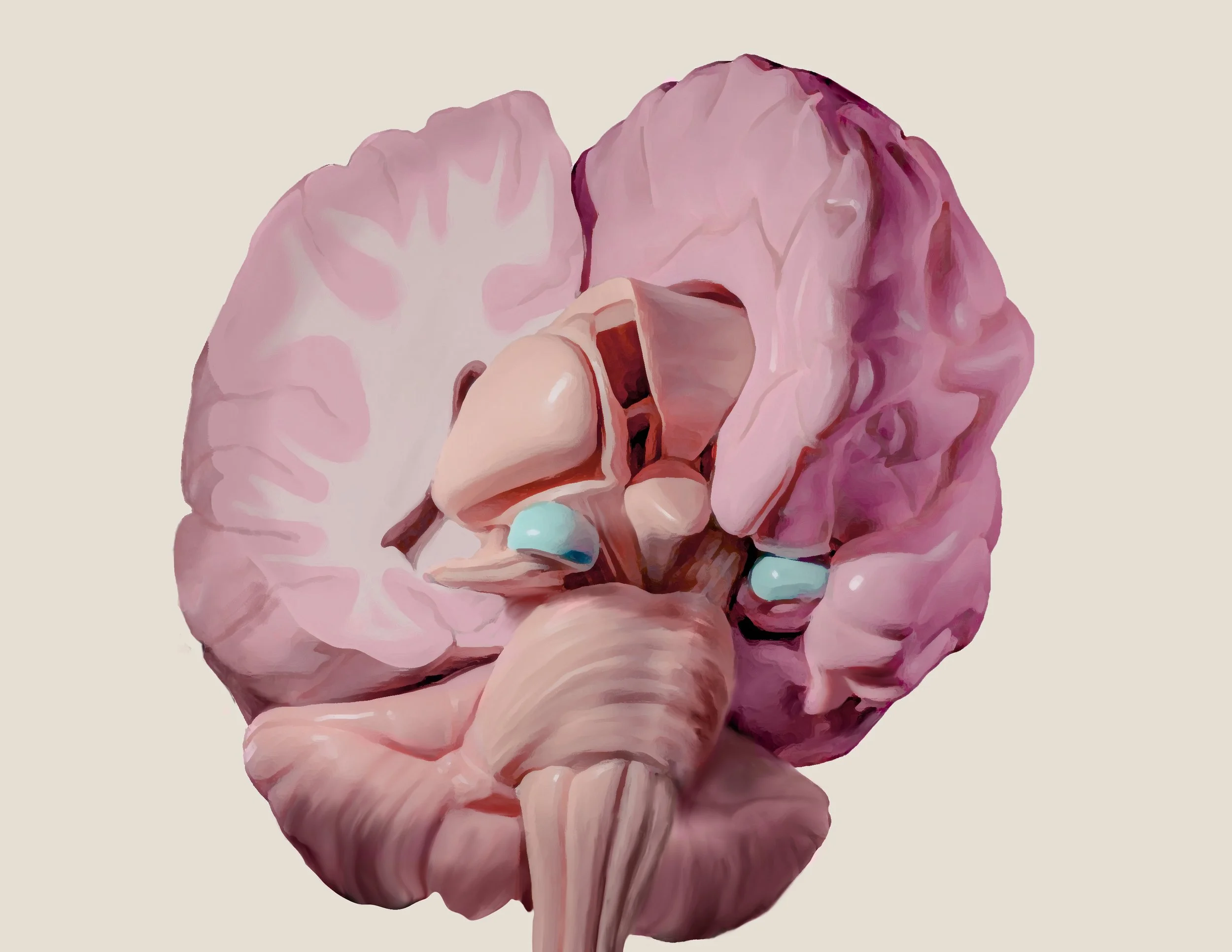
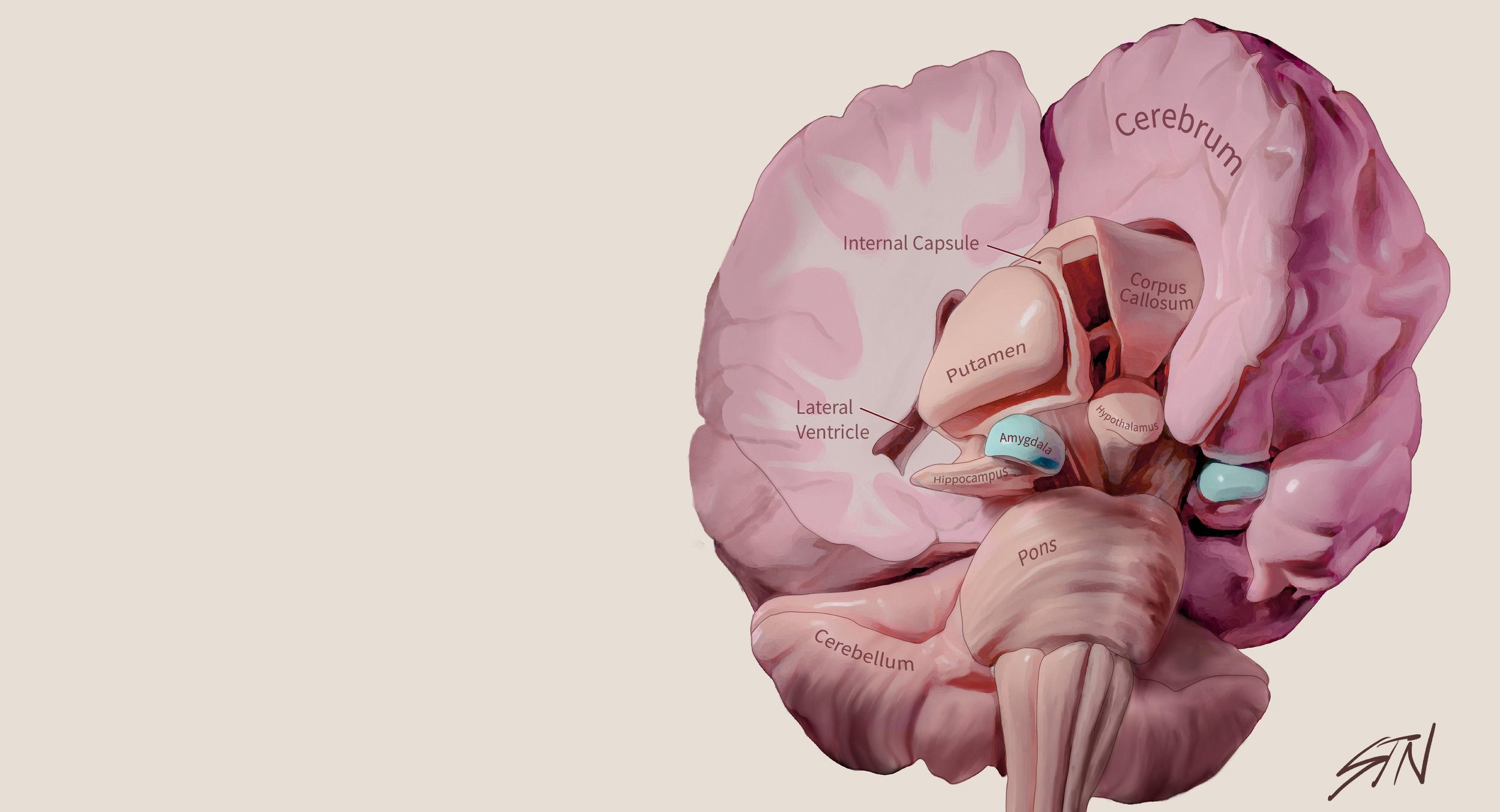
The goal of the project was to create an illustration to emphasize the spatial relationships of structures in the brain. The amygdala was chosen due to its complex function in the brain and the lack of existing illustrations showing its position in the brain. This illustration was made using a 3D model of the brain.
Media
Blender 3D, Procreate, Illustrator
Audience
Anatomy Student
Client
Shelley Wall
Year
2022
Inspirations
PureRef was used to compile helpful brain illustrations and references for the project. Many different styles and formats of illustrations were collected to assure the accuracy of the illustrations. I found I was drawn to soft palettes, and more artistic renditions of the brain.

Sketches
Many topics were considered. I ended up choosing the amygdala due to a lack of other resources its spatial relationship to the rest of the brain.
I chose to do an inferior antero-lateral view in order to show the amygdala from a non-traditional angle. I also felt that the view allowed for the student to see the main components of the limbic system. I did a grayscale rendering to see how the lighting could look.


Revealing the Amygdala
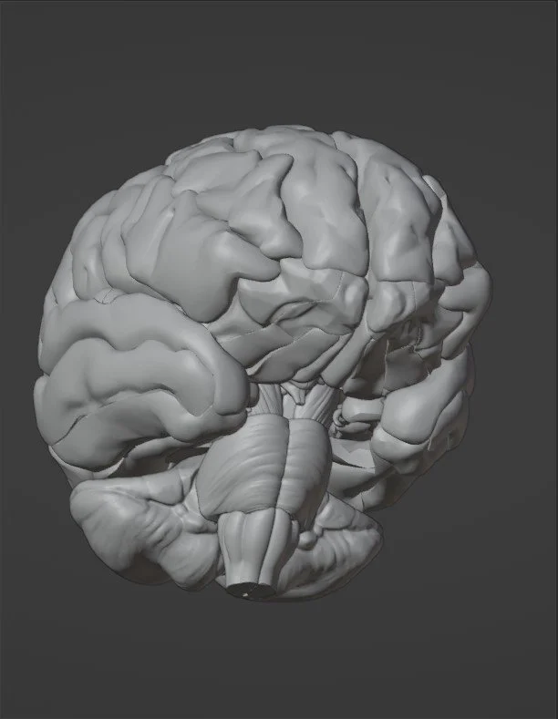
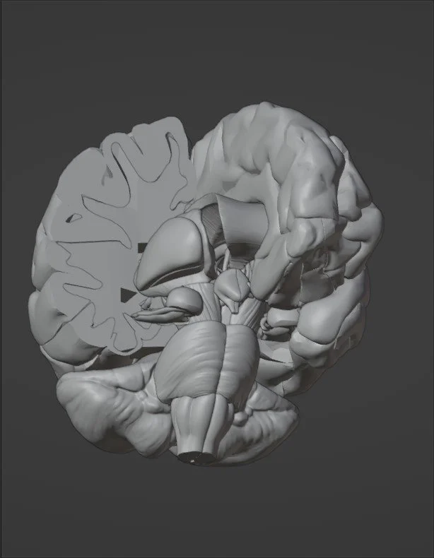
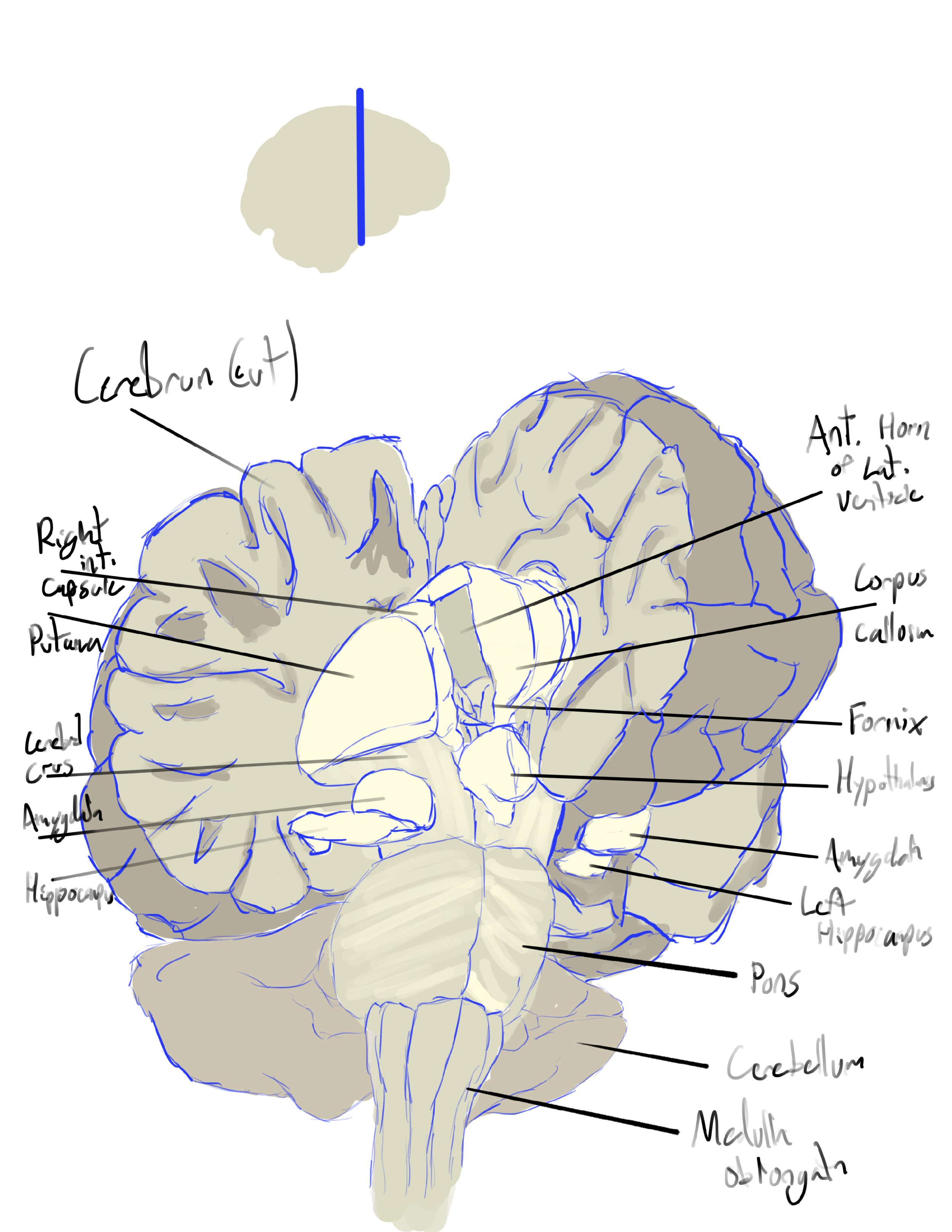
A full brain model was downloaded from BodyParts3D. At this point, the model was trimmed down to reveal the amygdala and start to find the final view. Many structures were hidden and a boolean was applied to reveal all the necessary structures and simulate a cut in the brain.


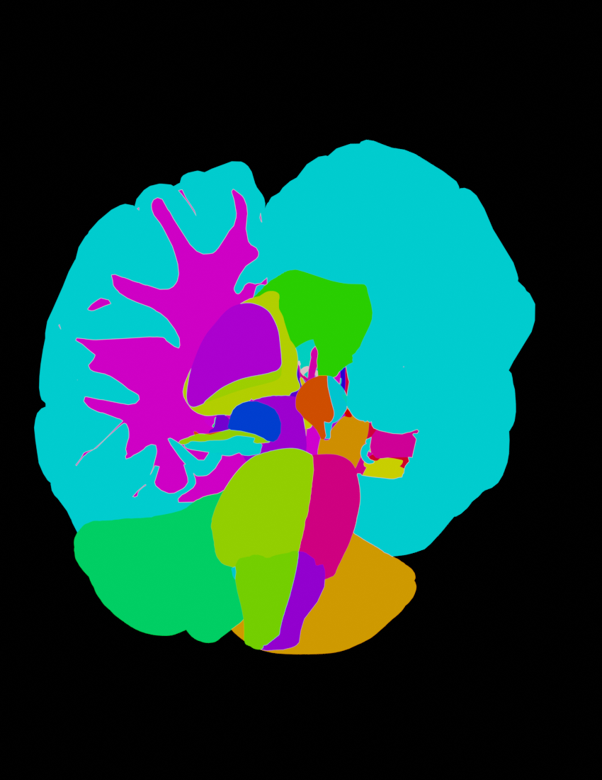
Rendering
Simplistic materials were used for color coding and light behavior.
The lighting used was a 3-point perspective lighting set up to show the 3D forms of the subject. The rim and backlights were colored slightly blue to cause cooler shadows for a painterly effect.
Two main renders were done. The first was a classical render of the scene. Orthographic view was used to preserve anatomical relations.
The second render was a pass of all the structures with different colors. This allowed for easy masking in Photoshop and procreate to create hard edges and isolate structures as needed.

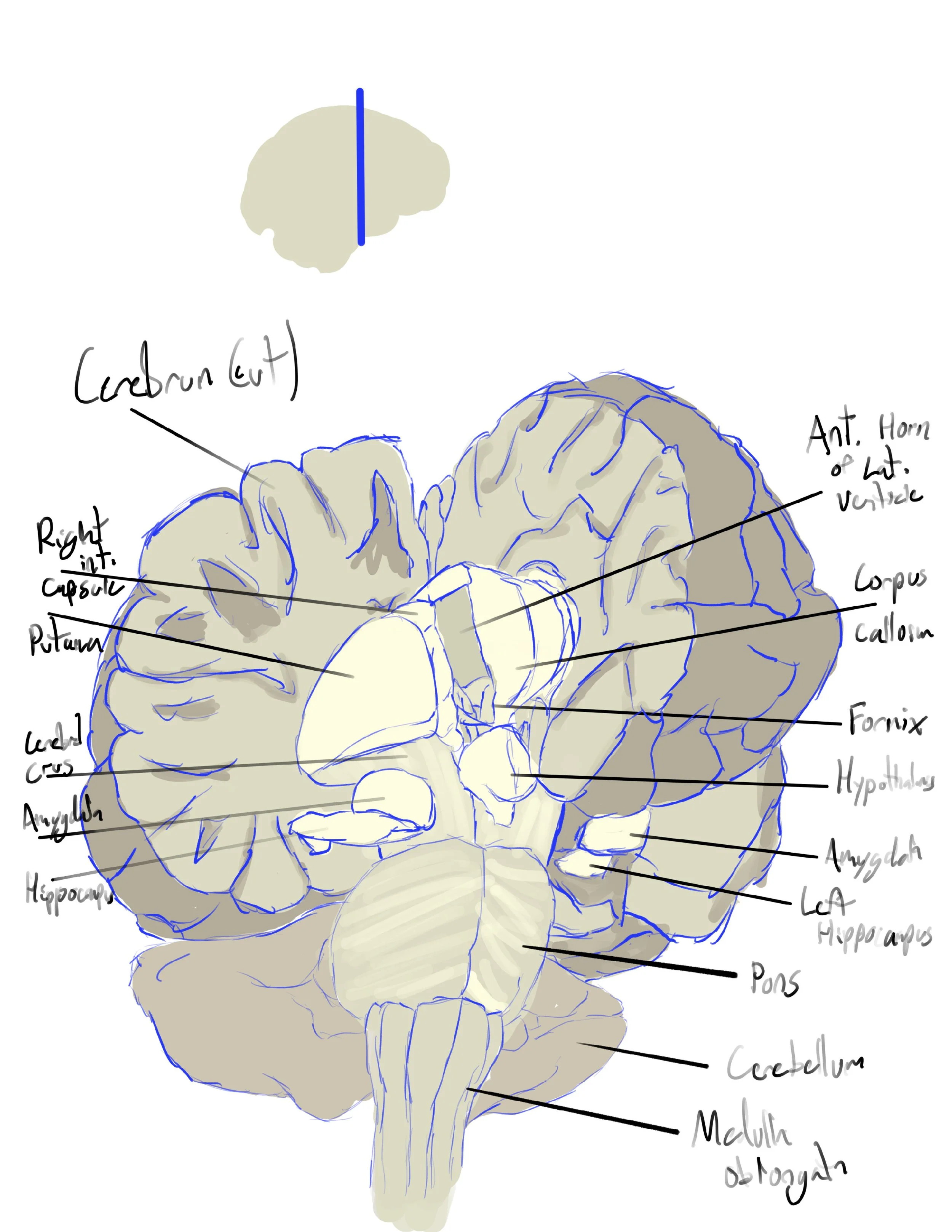
Painting and Labeling
The final render was then brought into procreate where it was painted and texture was added.
Many of the structures were adjusted here to be more anatomically accurate. As BodyParts3D is modular, there end up being odd shapes and cuts in many parts of the brain. These were painted over to make a more realistic brain.
The final painting was exported to Illustrator for labeling and adding titles. A vector orientation image was added.


References
Nieuwenhuys, R., Voogd, J., & van Huijzen, C. (2008). The human central nervous system. The Human Central Nervous System, 1–967. https://doi.org/10.1007/978-3-540-34686-9/COVER
Rubin Michael et al. (2007). Netter's Concise Neuroanatomy. Saunders Elsevier.
Venkatesh, A. (2016). Thieme Atlas of Anatomy, Volume 3: Head, Neck and Neuroanatomy, 2nd edn By M. Schuenke, E. Schulte, U. Schumacher; Consulting Editors: B. R. MacPherson, C. Stefan.
(ISBN 978‐1‐62623‐120‐7.) New York, Stuttgart, Delhi, Rio de Janeiro: Thieme Medical Publishers. 2016. Journal of Anatomy, 229(4), 600. https://doi.org/10.1111/JOA.12521
amygala surface modeling. (n.d.). Retrieved December 4, 2022, from https://pages.stat.wisc.edu/~mchung/research/amygdala/
Morphology of the Human Amygdala | Radiology Key. (n.d.). Retrieved December 4, 2022, from https://radiologykey.com/morphology-of-the-human-amygdala/#CR33
Saygin, Z. M., Osher, D. E., Augustinack, J., Fischl, B., & Gabrieli, J. D. E. (2011). Connectivity-based segmentation of human amygdala nuclei using probabilistic tractography. NeuroImage, 56(3), 1353. https://doi.org/10.1016/J.NEUROIMAGE.2011.03.006
Boccardi, M., Frisoni, G. B., Hare, R. D., Cavedo, E., Najt, P., Pievani, M., Rasser, P. E., Laakso, M. P., Aronen, H. J., Repo-Tiihonen, E., Vaurio, O., Thompson, P. M., & Tiihonen, J. (2011). Cortex and amygdala morphology in psychopathy. Psychiatry Research: Neuroimaging, 193(2), 85–92. https://doi.org/10.1016/J.PSCYCHRESNS.2010.12.013
Brabec, J., Rulseh, A., Hoyt, B., Vizek, M., Horinek, D., Hort, J., & Petrovicky, P. (2010). Volumetry of the human amygdala - an anatomical study. Psychiatry Research, 182(1), 67–72. https://doi.org/10.1016/J.PSCYCHRESNS.2009.11.005
Limbic System: Amygdala (Section 4, Chapter 6) Neuroscience Online: An Electronic Textbook for the Neurosciences | Department of Neurobiology and Anatomy - The University of Texas Medical School at Houston. (n.d.). Retrieved December 4, 2022, from https://nba.uth.tmc.edu/neuroscience/m/s4/chapter06.html
All Images